A team of researchers at Tufts caused tadpoles to process information using eyes grafted onto their tails after the tadpoles were treated with a human-approved migraine drug, according to Michael Levin, a professor in the Department of Biology and director of Tufts' Allen Discovery Center.
The original study, which Levin conducted alongside postdoctoral associate Douglas Blackiston and graduating senior Khanh Vien, was published online on March 30 in Nature Partner Journals' Regenerative Medicine section.
According to Levin, the purpose of the study was to observe the plasticity of the brain and to make the transplanted organs on tadpoles more functional. After being treated with the drug, many of the tadpoles grew nerves connecting the eye implants to their spinal cords, Levin said.
The species used in the study was a large aquatic frog commonly known as the African clawed frog. According to Blackiston, their neural pathways and hormones are very similar to those of humans.
“A lot of what we learn about the brain and neural connections and these drugs behave almost exactly the same between the animals,” Blackiston said.
He explained that the blind tadpoles were created through surgical techniques in which their developing eye tissue was removed while they were still embryos. According to Levin, the eyes were then grafted onto the tails of the tadpoles through cut-and-paste surgical operations.
“When you add the eye, a very small percentage of them can see. What we wanted to do was to make it much more efficient -- we wanted more of the animals to be able to see, and we wanted them to be able to see better,” Levin said. “That eye tissue puts out new nerves, and those nerves connect to the spinal cord of the animal. So we asked, ‘Could we increase the amount of innervation that comes out?’”
According to Blackiston, in the initial study, the eyes were grafted on the tadpoles’ heads but were moved farther and farther back on the animals as a test. He was surprised that some of the tadpoles were able to see with eyes so far away from their original location.
“That’s when we moved on to this study where we screened a huge amount of drugs ... that are commonly available, and we zeroed in on this one that increased the amount of animals that can see with these eyes even on the tail,” Blackiston said.
According to Blackiston, the drug that they found the most success with in increasing the visual processing of the tadpoles’ eyes was Zolmitriptan, a common pharmaceutical taken for migraines. Vien, the student who developed a test of the tadpoles' eyesight, explained that it was important to test drugs that are already known to be safe to humans.
"We wanted to focus on drugs that were human-approved because that was the human application. You want something that has the potential to be translational as soon as possible,” Vien said. "Otherwise, novel compounds would take three years to get through FDA certification, and then it would have to go through a clinical trial.”
For Levin, one of the most interesting parts of this treatment is that only the new nerves were affected by the drug.
“The normal innervation of the animal and all the other organs that are in their correct location ... ignore the drug entirely and are perfectly normal," he said.
He added that this study was significant because it gives more insight into the brain's plasticity or its ability to change. According to Levin, this could lead to human treatments in the future.
“If we understood how [the plasticity of the brain] works, we can make all kinds of cool technology for sensory motor augmentation,” Levin said. “Meaning if somebody lost an arm, they could have an electronic arm, or a new sensory organ, and we could connect it to all sorts of external devices because the brain is highly plastic and it can learn to use things that it was not normally evolved to use.”
Blackiston said that another takeaway of the study was the actual connection of the structures to their host, which shows that there could be ways to improve the functionality of human implants through less-invasive procedures than surgery.
Levin also noted that the study shows potential to use other pharmaceuticals beyond their original purposes.
“There are all kinds of drugs that humans already take," he said. "All these drugs are a potentially rich toolkit of solutions for all kinds of problems and regenerative medicine."
According to Blackiston, the next step in this line of research is figuring out how these eyes are connecting to the hosts. As of now, Blackiston explained that instead of connecting directly to the brain, they seem to be connecting to the spinal cord.
“We have a number of high-powered techniques that can trace neural connections, and we’re going to get down into the details of which ones are necessary, how those are formed and how those neurons talk to each other," he said.
Levin said that the team plans to take this research beyond eyes to continue testing the plasticity of the brain on other sensory organs and limbs.
“We have a collaboration with a team that ... is trying to do eye transplants in humans. So we’re going to be working with them to see if we can push this into a medical application,” he explained. “We’re going to work more on the basic question of how these cells figure out that they’re in the wrong place [and] how they know where they’re located in the body.”
Tufts researchers discover way to improve eye implants in tadpoles
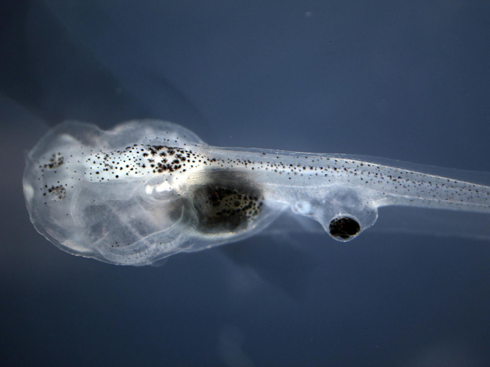
Blind tadpoles with eyes grafted onto their tails were able to process visual information after being treated with a small molecule neurotransmitter drug.